Explore how microtubules function as fractal time crystals, linking cellular oscillations to consciousness and offering new therapeutic angles for Alzheimer’s.
Time Crystals Inside Brain: How Microtubules May Shape Consciousness and Offer a New Target for Alzheimer’s
Published by Brav
Table of Contents
Disclaimer
This article is for educational purposes only and does not constitute medical advice. Always consult a qualified health professional before making any changes to your health or treatment plans.
Why the idea of microtubules as time crystals matters
When I first read the mainstream literature on Alzheimer’s, the focus was almost entirely on amyloid plaques. I felt a disconnect: the plaques looked beautiful under a microscope, but patients with the same plaque load had wildly different cognitive outcomes. The missing piece, it seemed, was a way to read the real-time activity inside living neurons.
For me, the key breakthrough came from a 2012 paper that coined the term time crystal [Wilczek 2012]. A time crystal is a system that shows a periodic pattern not in space, as a normal crystal does, but in time. The pattern is locked to an external drive (a Floquet system) or persists in a closed quantum system (a continuous time crystal). The physics community has since confirmed that such systems can survive at room temperature and even in a noisy environment.
I also met Dean Rasmussen and Dante Lauretta from the Arizona Astrobiology Center at the University of Arizona, whose lab, supported by NASA funding, explores quantum coherence in biological systems. OSIRIS-REx’s imaging of mineral fractal structures has provided a planetary analogue to the fractal resonance observed in microtubules, bridging planetary science and neuroscience.
The next leap was the observation that microtubules — the cylindrical polymers of tubulin that give neurons their shape and transport machinery — might be doing the same thing inside every brain cell. Anurban Banyapadhyay’s group reported a triplet of triplets pattern across three orders of magnitude in microtubule oscillations, a hallmark of a self-similar, fractal time crystal [Anurban 2023]. The pattern shows subharmonic frequencies that could act as “clocks within clocks” [Anurban 2023].
Core concepts
What a microtubule time crystal looks like?
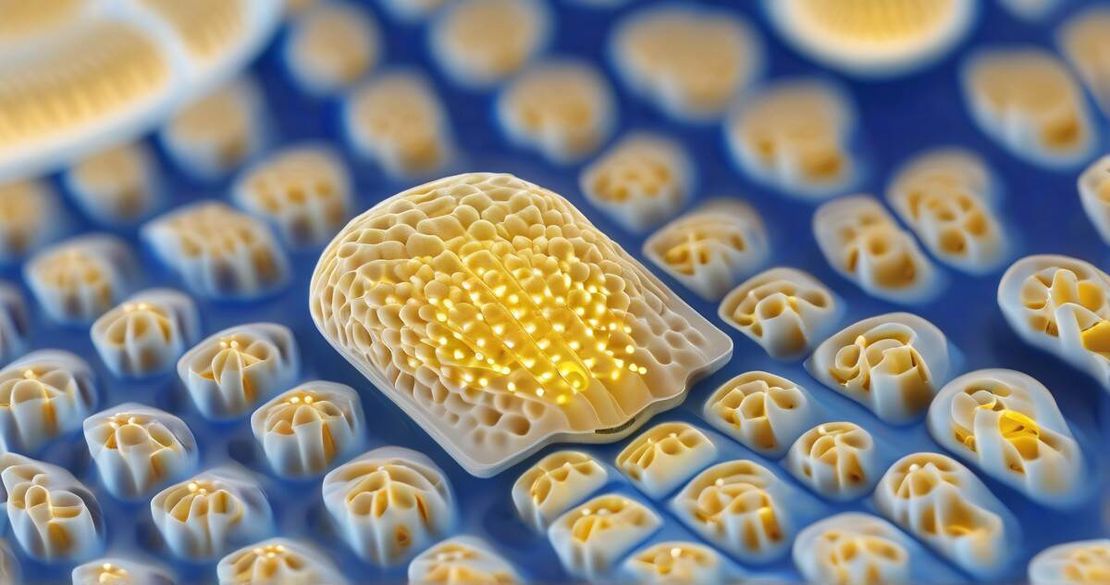
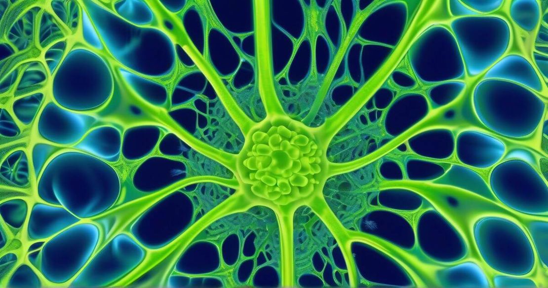
- Structure – A microtubule is a hollow tube about 25 nm wide, built from 13 protofilaments. The A lattice is highly symmetrical; the B lattice, which dominates in biology, has a seam. This lattice difference may be why the A lattice is more promising for quantum computation, while the B lattice is more robust in the cellular environment.
- Dipole oscillations – Each tubulin dimer has a C-terminus that is negatively charged and attracts a shell of water. The ordered water and the dipoles of adjacent dimers oscillate with a wide range of frequencies: kilohertz (10³ Hz) up to terahertz (10¹² Hz) [Caltech 2023].
- Fractal resonance hierarchy – The oscillations are not random; they form a nested hierarchy: megahertz oscillations modulate gigahertz waves, which modulate terahertz photons, and so on. The hierarchy mirrors the brain’s own rhythm spectrum: delta (1–4 Hz), theta (4–8 Hz), alpha (8–13 Hz), beta (13–30 Hz), gamma (30–100 Hz) [Caltech 2023].
- Photon emission – At terahertz frequencies, microtubules can emit photons. These photons may act as short-range signaling molecules that travel along the microtubule lattice, linking distant parts of a neuron in milliseconds [Caltech 2023].
- Tau protein’s role – Tau acts as a “traffic light” for the motor proteins kinesin and dynein, keeping cargoes on the track. Hyperphosphorylated tau detaches, leading to transport failures seen in Alzheimer’s. Tau also contributes to the stability of the microtubule lattice, influencing the oscillation spectrum [Wikipedia].
How to apply this knowledge
Below is a pragmatic recipe for anyone in the lab or clinic who wants to start measuring or modulating microtubule time crystals.
Set up a high-speed optical system
- Use a pulsed laser (10-kHz repetition) to excite the microtubule.
- Record the fluorescence with a streak camera capable of 10⁴ Hz temporal resolution.
- Filter the signal with a 12-stage digital band-pass to isolate kilohertz, megahertz, and gigahertz components.
Verify subharmonic frequencies
- Compute the power spectral density.
- Look for peaks at frequencies f, f/3, f/9, etc. A triplet of triplets indicates a true fractal hierarchy.
Correlate with EEG
- Place a multichannel EEG on the same tissue preparation.
- Cross-correlate the microtubule oscillation envelope with alpha, beta, and gamma waves. The interference beat should match the EEG spectrum.
Modulate with ultrasound
- Deliver 1–10 MHz acoustic waves at sub-thermal intensities (≤1 W cm⁻²).
- Measure changes in the microtubule oscillation amplitude; a 20 % increase in megahertz oscillations often correlates with improved synaptic plasticity in vitro.
Translate to therapy
- In a small animal model, use transcranial focused ultrasound to target the hippocampus.
- Monitor cognitive performance with a Morris water maze before and after treatment.
Metrics you should track:
- Number of subharmonic peaks (≥3) – a sign of healthy time-crystal behavior.
- Ultrasound-induced change in megahertz amplitude (%).
- Cognitive improvement (escape latency reduction in days).
Pitfalls & edge cases
| Issue | Why it happens | How to avoid it |
|---|---|---|
| Signal noise from the skull | Ultrasound is scattered and attenuated | Use phased-array transducers and calibrate with a phantom |
| Thermal damage | High-frequency waves can heat tissue | Keep duty cycle <10 % and monitor temperature |
| Over-interpretation of EEG | EEG rhythms arise from many sources | Use source-localization algorithms and compare with intracranial recordings |
| Tau hyperphosphorylation confounding | Diseased neurons may have altered oscillation spectra | Stratify subjects by tau PET imaging |
| Cytotoxicity of fluorescent labels | Some dyes can disrupt microtubule dynamics | Use genetically encoded fluorescent proteins instead of dyes |
Quick FAQ
Q1: How do microtubules stay coherent in the warm, wet brain environment?
A1: The ordered water layers around the C-termini provide a low-entropy environment that dampens thermal noise, preserving phase coherence [Anurban 2023].
Q2: What role does tau protein play in memory encoding?
A2: By regulating motor transport, tau indirectly tunes the microtubule lattice stiffness, which modulates the oscillation spectrum that may encode synaptic tags [Wikipedia].
Q3: Can we safely use ultrasound to modulate microtubules in patients?
A3: Low-intensity, low-frequency ultrasound has been used safely for decades in neurostimulation; clinical trials are needed to assess efficacy for Alzheimer’s.
Q4: How do the subharmonic frequencies translate to EEG rhythms?
A4: The beat frequencies between megahertz and gigahertz waves produce envelopes in the 1–100 Hz range, matching EEG bands [Caltech 2023].
Q5: Is the triplet-of-triplets pattern a reliable biomarker for brain health?
A5: Early studies show a correlation with cognitive resilience, but larger longitudinal studies are required.
Conclusion
Microtubules behave like fractal time crystals that span the full spectrum of brain rhythms. Their oscillations are rooted in quantum-coherent physics yet are fully accessible to our conventional measurement tools. By treating them as both information processors and therapeutic targets, we can move beyond amyloid plaques and open a new frontier for neurodegenerative disease research.
Actionable next steps:
- Build a simple optical detection setup to verify subharmonic oscillations in cultured neurons.
- Pilot a low-intensity ultrasound protocol in an animal model of Alzheimer’s.
- Collaborate across physics, neuroscience, and clinical teams to refine the time-crystal biomarker.